Cardiac Arrhythmias in Horses
Clinical Connections – Summer 2023
Jenny Reed, Lecturer in Equine Medicine
Equine arrhythmias result in a range of effects, from poor performance and reduced exercise capacity to sudden cardiac death – but are also present in healthy equine athletes.
On identifying an arrhythmia, the veterinarian’s role is to determine whether a physiological or pathological arrhythmia exists and to assess the risk to the rider. Unfortunately, this is often difficult, and whilst auscultation is useful, a definitive diagnosis is usually achieved via electrocardiography (ECG).
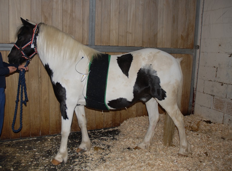
The introduction of smartphone-based ECG technology has allowed for the diagnosis of rhythm disturbances at rest and under field conditions [1, 2], however the identification of arrhythmias occurring at exercise requires a telemetric system with detailed analysis of the ECG following collection – a service offered at RVC Equine.

Exercising ECG recordings are difficult to analyse due to the presence of motion artefacts and the lack of P-waves and short QRS intervals associated with physiological tachycardia [3]. Whilst morphological changes may be identified on subjective analysis, heart rate variability has more recently been utilised in the detection of arrhythmias in horses at exercise [4], and aids in interpretation of ECGs notwithstanding the aforementioned challenges.
Heart rate variability, the difference in heart rate between consecutive cycles, is a normal phenomenon related to changes in the autonomic nervous system. However, abnormal variations in heart rate during exercise suggest premature or late beats which are identified by computer-aided evaluation of beat-beat intervals. Despite the advancement of technology aiding in collection and analysis of ECG’s, characterisation of arrhythmias remains a challenge.
The presence of morphological and/or temporal changes in the ECG can be suggestive of the source of an ectopic beat, however occasionally these findings do not agree to the source, and as a result a more descriptive approach to arrhythmias has been suggested [5].
While ECG analysis may help to locate the likely source of an arrhythmia, catheter-based intra-cardiac electroanatomic mapping is being used to provide information on the precise location of electrophysiological abnormalities. This involves the placement of an intra-cardiac steerable catheter and the creation of a magnetic field over the heart via which the location of the catheter is ‘visualised’.
By combining both 3D anatomical and electrophysiological data, an electrical map of the inner surface of the heart is created. Initial studies utilised this 3D mapping system to describe the normal activation pattern of the myocardium throughout the cardiac cycle, and while initially performed in anaesthetised horses, the technology has also been used in standing horses [6].
In addition to demonstrating the normal propagation of the electrical impulse through the heart, the depolarisation of the ventricular myocardium was also shown to be reflected in the QRS morphology, and subsequently, 12-lead ECG’s have also been evaluated as a method for identifying the origin of focal premature atrial and ventricular depolarisations [7,8].
The ability to detect the source of ectopy also allows for the treatment of arrhythmias with intra-cardiac radiofrequency ablation. Several atrial arrhythmias have been successfully treated in this way, including atrial tachycardia [9] and an atrioventricular accessory pathway [10].
Whilst these technologies are not universally available, one catheter-based treatment that has been performed at the RVC Equine Hospital is trans-venous electro-conversion (TVEC). Atrial fibrillation (AF) is the most common arrhythmia affecting performance in horses, and conversion to sinus rhythm can be accomplished via several methods. Quinidine sulphate is effective for pharmacological conversion of atrial fibrillation in horses, but undesirable side effects are frequently encountered. A general anaesthesia is required for TVEC, however both methods have high success rates and recurrence rates are similar.
While it remains a constantly evolving field, our ability to more accurately detect, describe and locate equine arrhythmias has improved with the introduction of these technologies, and the opportunities for successful resolution of arrhythmias continue to grow.
References
1. Welch-Huston, B., Durward-Akhurst, S., Norton, E., Ellingson, L., Rendahl, A. and McCue, M. (2020), Comparison between smartphone electrocardiography and standard three-lead base apex electrocardiography in healthy horses. Veterinary Record, 187: e70-e70.
2. Alberti E, Stucchi L, Pesce V, Stancari G, Ferro E, Ferrucci F, Zucca E. Evaluation of a smartphone-based electrocardiogram device accuracy in field and in hospital conditions in horses. Vet Rec Open. 2020 Dec 21;7(1)
3. van Loon, G. (2022). Diagnosis cardiac arrhythmias in performance horses. Equine Exercise Physiology, 11th International Conference, Abstracts. Presented at the International Conference on Equine Exercise Physiology (ICEEP), Uppsala, Sweden.
4. Frick, L, Schwarzwald, CC, Mitchell, KJ. The use of heart rate variability analysis to detect arrhythmias in horses undergoing a standard treadmill exercise test. J Vet Intern Med. 2019; 33: 212– 224.
5. Slack, J., Stefanovski, D., Madsen, T. F., Fjordbakk, C. T., Strand, E., & Fintl, C. (2021). Cardiac arrhythmias in poorly performing Standardbred and Norwegian-Swedish Coldblooded trotters undergoing high-speed treadmill testing. Veterinary journal (London, England : 1997), 267, 105574.
6. Hesselkilde, E, Linz, D, Saljic, A, et al. First catheter-based high-density endocardial 3D electroanatomical mapping of the right atrium in standing horses. Equine Vet J. 2021; 53: 186– 193.
7. Van Steenkiste, G., Delhaas, T., Hermans, B., Vera, L., Decloedt, A., & van Loon, G. (2022). An Exploratory Study on Vectorcardiographic Identification of the Site of Origin of Focally Induced Premature Depolarizations in Horses, Part I: The Atria. Animals: an open access journal from MDPI, 12(5), 549.
8. Van Steenkiste, G., Delhaas, T., Hermans, B., Vera, L., Decloedt, A., & van Loon, G. (2022). An Exploratory Study on Vectorcardiographic Identification of the Site of Origin of Focally Induced Premature Depolarizations in Horses, Part II: The Ventricles. Animals: an open access journal from MDPI, 12(5), 550.
9. Van Steenkiste, G, Boussy, T, Duytschaever, M, et al. Detection of the origin of atrial tachycardia by 3D electro-anatomical mapping and treatment by radiofrequency catheter ablation in horses. J Vet Intern Med. 2022; 36( 4): 1481- 1490.
10. Buschmann, E, Van Steenkiste, G, Boussy, T, et al. Three-dimensional electro-anatomical mapping and radiofrequency ablation as a novel treatment for atrioventricular accessory pathway in a horse: A case report. J Vet Intern Med. 2023; 37( 2): 728- 734.
