Cardiothoracic Surgery Team Progress
Clinical Connections – Summer 2024
Dan Brockman, who leads the team and is Professor of Small Animal Surgery, outlines developments in the Cardiothoracic Surgery Service, which are enabling more dogs to be treated. The RVC’s service, which was established in 2005, is the only place in the UK offering “open-heart” surgery to canine patients. As Dan explains, other elements of the team, in addition to the surgeons, are vital to deliver an exceptional service to dogs and their owners.
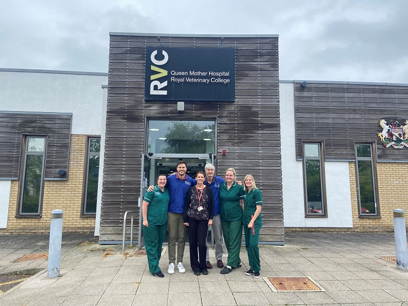
In common with many initiatives, we lost a bit of ground due to the impact of COVID in recent years but we are making it up again now. Our team is strengthening and we’ve recently hired two Registered Veterinary Nurses; Rachel Collin and Kayley Dowd, who are dedicated to the cardiothoracic service both of whom have extensive previous experience in busy practices.
They are working alongside Sarah Carey, our Cardiothoracic Service Co-ordinator, and have been learning the ropes very quicky. Our nurses are crucial to the success of the programme and have a very broad ranging role – they are patient co-ordinators, theatre nurses, deliver ICU care and support general client communication and facilitation of visits. It’s a slightly different role from most traditional nursing roles in the hospital as it’s not discipline-specific – they have to cross lots of disciplines.
The nurses meet the clients and follow the patients all the way through their care. The people who manage this particular group of canine patients need to understand the needs of the dogs which are subtly different from many other patients. The consistency of having the same person look at the animal multiple times a day, allows them to pick up very subtle changes – and those subtle changes can be very important!
Having a core group of people who work with these patients all the time, rather than the patient moving from ward to ward, where a new group of people have to get familiar with them, enables better consistency of care – and therefore also better consistency of results.
That continuity of care is also ideal for interaction with clients – they love knowing that they will see a certain nurse and a certain vet. Because it’s often such an emotional experience for owners, they really like the consistency of seeing a familiar face every time they come to the hospital and hearing a consistent voice on the phone. The client care aspect of cardiothoracic surgery is so important, as these pet owners are often extremely emotionally bonded clients.


Surgical team developments
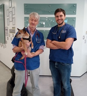
Matteo Rossanese [Lecturer in Soft Tissue Surgery] declared an interest in open heart surgery some years ago and has been “scrubbing in” with me for a couple of years. Matteo is a very gifted surgeon who has already completed advanced surgical training and so having such a competent pair of hands involved with the surgery is extremely helpful.
We’ve got to the stage now where we essentially assist each other – we take it in turns to be the primary surgeon and assistant surgeon. Matteo is acting as the primary surgeon with guidance now and I would hope that, given another year or so, he will be capable of doing the procedure on his own, without my input.
We are also hoping to get both a Cardiothoracic Surgery Fellow and Perfusion Fellow back on the books this year. It’s about succession planning for me now – I’d ultimately like to walk away from this programme by the time I retire knowing that it’s sustainable and it will continue. The demand is enormous but the supply worldwide of people who can do mitral valve repair is low and we have an important part to play in building more capacity. We are booking cases into 2025 already.
We are currently doing two surgeries a week again, which is what we were doing before COVID, and if we can attract more anaesthesiology and cardiology personnel, we can potentially go up to three cases a week. It becomes much easier to train people when you have a higher throughput as the trainee will “climb” the “learning curve” more quickly.
In relation to the broader team, we have two new cardiologists [see spring’s issue of Clinical Connections], Nekesa Morey and Joshua Hannabuss, and they, along with our enthusiastic team of cardiology residents, are extremely helpful to the programme.
Technology
In addition to human expertise, technology is critical to what we do. We are fortunate to have a brand new cardiopulmonary bypass console (heart-lung machine), which was purchased through donations to the RVC Animal Care Trust that were restricted for use in the development of heart surgery. This is the machine that circulates the blood once the heart is bypassed and we have it open.
The ACT also provided funds to purchase surgical loupes for Matteo. These are an essential piece of equipment but they are extremely expensive so we are very grateful to the RVC’s Animal Care Trust and donors supporting our work.
Amber’s story
Last September Amber, a 10-year-old Yorkshire terrier cross, came for evaluation by the Cardiology Service and myxomatous mitral valve disease was confirmed. The possibility of being a candidate for mitral valve repair was discussed.
Amber's heart disease was stage C, meaning her heart was enlarged secondary to her valvular disease and that resulted in her developing congestive heart failure. Her valve was severely affected and there was evidence of ruptured chords, which may have been responsible for a collapse weeks before.
Amber returned to the RVC at the end of October for mitral valve repair, under cardiopulmonary bypass. A left 5th intercostal thoracotomy was used to access the heart and a small approach to the left cervical region to isolate the carotid artery for cannulation was made. The mitral valve was accessed via a left atriotomy under conditions of cardiopulmonary bypass.
Amber’s mitral valve was severely diseased with thickening to all valve edges, stretching of all chords and a small fissure in the parietal valve leaflet.
Three artificial chords (Gore-Tex sutures) were placed between the papillary muscles and the edge of the septal valve leaflets (CV 6), and two artificial chords were placed in the P2 valve leaflet (CV 6). The fissure was closed with a single suture of 6-0 prolene. In addition, we performed a mitral purse-string annuloplasty with pledgeted CV5 Gore-Tex suture to bring the orifice where the valve sits back down to a normal size.
Amber progressed very well after surgery and echocardiography confirmed that the repair was a success. She was discharged from hospital on November 6th.
A month after surgery Amber came in for post mitral valve repair evaluation. Her energy levels had returned and her appetite had improved. Echocardiography confirmed that the valve repair was stable, there was trivial residual mitral valve regurgitation and no signs of clots or thrombi. The echo measurements confirmed that Amber's heart had returned to a normal size.
Amber was also examined at the RVC for her three-month post mitral valve repair evaluation and this confirmed that the repair was stable and her heart was functioning normally. Amber was doing extremely well, off all medications and enjoying life to the full.
