Deaf Dog Has Grass Seed Behind Eye Removed
Clinical Connections – Autumn 2022
A year-old cocker spaniel presented to the Emergency and Critical Care service in July for acute onset redness and swelling of the left eye, which had started two days before. Marvin had been running through tall grass and his local vet removed several grass seeds from his mouth.

Marvin has been deaf since birth and is missing a toe pad but was always lively and playful with another dog in the household until his eye deteriorated.
Clinical signs were relatively mild on his first admission, when he was also seen by the Ophthalmology Service, so we were not able to justify further investigation and symptomatic treatment was given. However, clinical signs returned with a vengeance two weeks after stopping medication and he was readmitted in August.
During his first admission, Marvin was found to have some exophthalmos ocular discharge. He was sedated to have his eye thoroughly checked for any foreign material. A small puncture wound on the soft palate at the back of the mouth (left pterygopalatine fossa region) was found, which was draining bloody material.
When examined the next morning by the Ophthalmology Service there was no longer any protrusion of the eyeball. Marvin appeared comfortable and was holding the eye open, with only mild swelling of the conjunctiva. He could open his mouth fully, with no evidence of discomfort, so he was discharged from the hospital.
Foreign material may penetrate the soft part of the mouth but not become embedded or enter the tissue and remain there, where they then cause an abscess. Grass seeds can move in one direction but not the reverse, due to the presence of barbs. As Marvin's signs were subtle and he was comfortable and able to open his mouth easily, the team was less concerned about a retrobulbar abscess at that point.
From the description of his examination findings both by our Emergency Referrals service and the referring vet, it appeared that Marvin was responding to the antibiotic and anti-inflammatory injections that he received prior to referral. Therefore, rather than proceed with 3D imaging of the head to look for foreign material at that point, he was treated symptomatically with antibiotics and anti-inflammatories.
Deterioration and second admission

In August his owner noticed reddening in Marvin's eye, along with protrusion of the third eyelid. She administered some Celluvisc but over the next few days the protrusion increased and the eye closed. His local vet observed that the left side of Marvin's head was swollen. He was given oral anti-inflammatories, and, during the night, Marvin developed mucopurulent ocular discharge. No obvious discomfort in the mouth or discharge from the mouth was noticed but Marvin had become withdrawn and had stopped playing with the other family dog.
Upon return to the QMHA, the left eye was moderately blepharospastic with a markedly increased blink rate. There was mild mucopurulent discharge and the globe appeared medially displaced. There was ventral periocular swelling, which was hot on palpation and painful. The palpebral and bulbar conjunctiva was markedly hyperaemic and congested with severe chemosis of the lateral palpebral conjunctiva. There were mild signs of uveitis. Given the deterioration in clinical signs, further investigation was recommended.
CT identified a large swelling on the left side of the face, extending to the left masseter, consistent with an abscess. Although no clear foreign body was identified, that could not rule out a small unobserved object from being present. Ocular ultrasound can complement abnormalities seen on CT imaging. The radiologists identified a dorsal lateral subcutaneous abscess with grass seed, which was removed with ultrasound guidance under general anaesthesia. The area was explored and lavaged and left as an 'open wound' to heal by second intention.
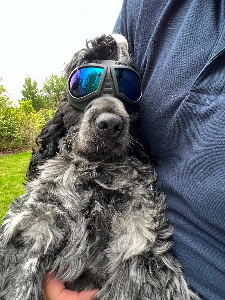
The following morning, Marvin showed a marked improvement in comfort and swelling so was discharged with a course of anti-inflammatories and antibiotics. Marvin is back to his normal self and has completely recovered. His owners are now trying to encourage him to wear goggles when out on walks to prevent a recurrence.
