World-First Surgery Combination Saves Puppy’s Life
Surgeons at the RVC have successfully carried out the surgical repair of a complex combination of heart defects in a dog. The abnormalities required tricuspid valve repair surgery along with the repair for the common atrium. It is the first time these procedures have been carried out on a dog in the same operation.
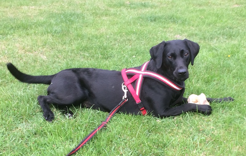
Lottie, an 11-month-old Labrador puppy appeared full of life with no problems until her owners took her to the local vets to be spayed. During a routine check before this procedure a very loud heart murmur was heard. A heart ultrasound revealed that Lottie had several defects in her heart, which she had been born with (congenital).

The two major ones were a malformation of her tricuspid valve, which is the valve that sits on the right side of the heart to regulate flow between the right filling chamber (atrium) and pumping chamber (ventricle), and the second a very large defect between the left and right filling chambers or atria, known as a “common atrium”.
Lottie was referred to the RVC’s cardiothoracic department for further evaluation and to see if there was anything that could be done to help her. The team, which is led by Dan Brockman, Professor of Small Animal Surgery, is notable for performing several cutting-edge surgeries, including a world-first treatment to save the life of a dog born with a malformed tricuspid valve.
Lottie underwent further heart ultrasound using 3D technology as well as a CT scan. Repair of the tricuspid valve has only been performed a handful of times and has not been done at the same time as repair of a common atrium.
Lottie’s owners decided to proceed with a surgical correction in order to try and help extend Lottie’s otherwise limited life and to preserve a good quality of life. This operation was undertaken on July 30th. Lottie had her heart stopped to perform the complex repair and her circulation to the rest of the body was maintained with the use of a heart lung machine run by a perfusionist from Great Ormond Street Hospital (Nigel Cross).
Commenting on the surgery and the number of different practitioners involved to help Lottie, Poppy Bristow, Fellow in Cardiothoracic Surgery at the RVC, said: “Altogether 10 people were involved in her operation and many more for her care before and after surgery, including veterinary specialists, veterinary nurses and veterinary specialists-in-training from surgery, cardiology, anaesthesia and emergency and critical care, as well as Lottie’s referring cardiologist and her local veterinary practice.
“Lottie’s heart was stopped for an hour and a half, with the whole operation taking four hours. Her malformed tricuspid valve was released by cutting its abnormal attachments and artificial chords using Gore-Tex material were placed. Her single atrium was then divided into two using a large patch of Gore-Tex. Lottie has made a good recovery so far and was walking around and eating from the day after her surgery. She was discharged back to her owners after six days and has continued to thrive at home.”
Professor Brockman added: “In Lottie, we had a young energetic dog with such a serious and life-limiting heart condition, that we were desperate to try and help her. The repair was complex but incorporated a combination of surgical manoeuvres that we had done before. With careful pre-operative planning and using our previous experience, we were able to design and execute the surgical treatment. It is still ‘early days’ but the initial signs suggest that Lottie is going to enjoy an excellent quality of life, following this operation and, we all hope, a normal lifespan.”

