Emergency contact: 01707 666399
Mitral Valve Transcatheter Edge-to-Edge Repair (M-TEER)
Degenerative mitral valve disease in dogs
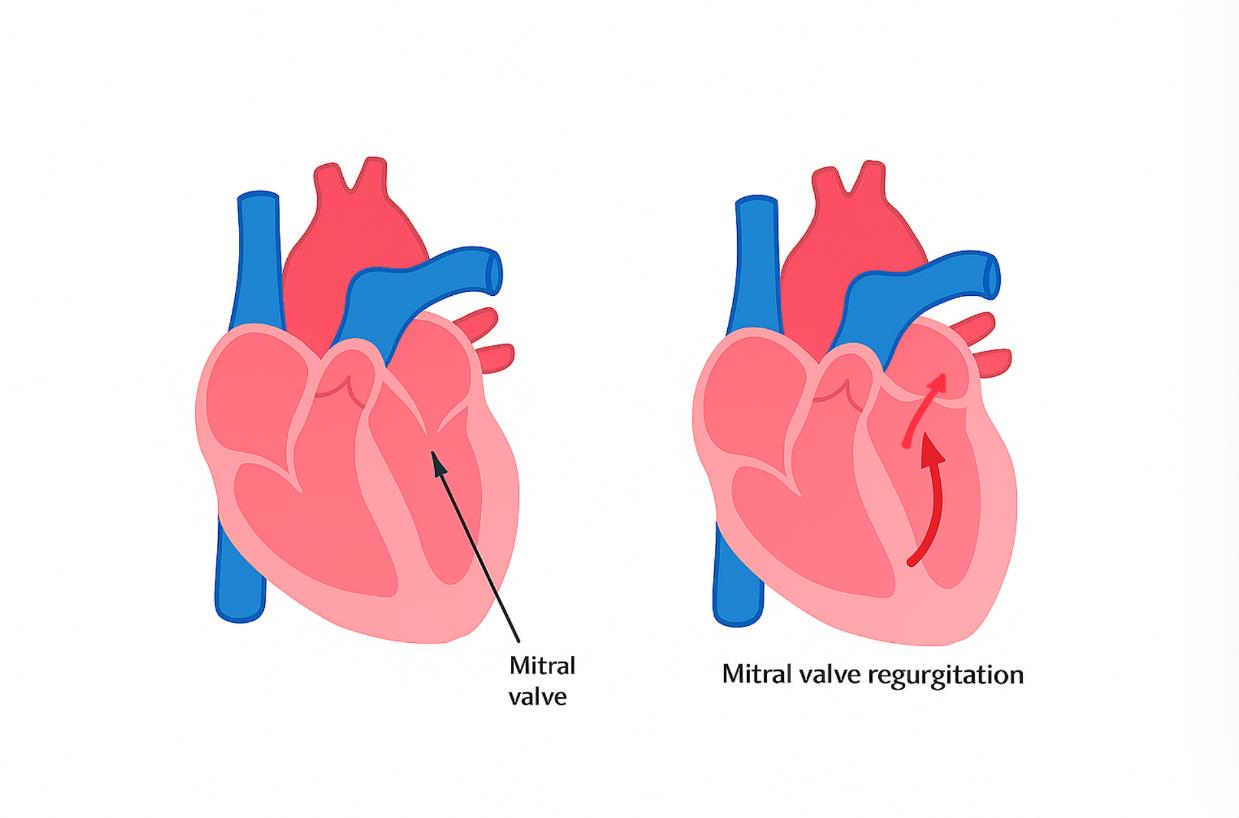
Degenerative mitral valve disease (DMVD), also known as myxomatous mitral valve disease (MMVD), is the most common heart disease in dogs, affecting predominantly small and medium sized breeds. It is characterized by abnormal changes to the mitral valve, resulting in regurgitation (leaking) through this valve. Over time, the left side of the heart may enlarge, which can eventually result in complications such as left-sided congestive heart failure (accumulation of fluid within the lungs) and difficulties breathing.
Treatment options for degenerative mitral valve disease
For dogs with mild degenerative valve disease and normal heart size (ACVIM stage B1), no treatment is required, but routine monitoring is advised. For dogs with moderate to severe degenerative valve disease resulting in heart enlargement (ACVIM stage B2), medical therapy with pimobendan (Vetmedin®) has been proven to delay the onset of clinical signs and delay the development of congestive heart failure (CHF). If a dog has advanced disease and has developed congestive heart failure (ACVIM stage C), additional medical therapies will be required. The goal of medical therapy is to maintain good quality of life for as long as possible, however, most of these dogs will ultimately succumb to their disease. The only treatments available that directly address the cause of mitral regurgitation are:
- Open heart surgery under cardiopulmonary bypass, to repair the mitral valve
- Mitral valve transcatheter edge-to-edge repair (TEER)
The Royal Veterinary College (RVC) is the first centre worldwide offering all options for the treatment of degenerative mitral valve disease in dogs (summarised in table 1).
Table 1: Summary of the treatment options for advanced degenerative mitral valve disease in dogs
|
|
Medical treatment |
Trans-catheter edge-to-edge repair (TEER) |
Open-heart surgery |
|
Procedure risk |
Low complications may include dehydration, electrolyte abnormalities or kidney injury |
Low to moderate 95% survive to discharge. Major complications (5%) most likely to occur within 48 hours of procedure. |
Low to moderate 90-95% survival to discharge. Major complications most likely to occur during hospitalisation. |
|
Quality of life after treatment |
Average survival time in stage C is 9-12 months. When stable on medications, quality of life is good, although relapse of clinical signs is likely, requiring hospitalisation or at home medication adjustments. |
Most dogs continue pimobendan following surgery however many will have a dose reduction in their heart failure medication. Some may have these medications stopped. Quality of life tends to be good. |
Most dogs have heart failure medications stopped immediately after surgery. Many dogs will go on to have their pimobendan stopped. Most dogs have an excellent quality of life. |
|
Survival rate at 2-years |
15% |
80% |
90% |
|
Price estimate (10/2025) |
Pimobendan (Vetmedin) for a 10kg dog is roughly £78 per month |
£12,500 |
£24,200 |
Transcatheter edge-to-edge repair (TEER)
Transcatheter edge-to-edge repair (TEER) is a minimally invasive procedure used to treat degenerative mitral valve disease in dogs. Compared to open-heart surgery (requiring heart-lung bypass), TEER is a catheter-based technique performed via a small incision on the side of the chest resulting in a short period of hospitalisation. Whilst TEER is a new procedure in the UK, it has been available since 2020 in Asia and 2021 in the USA. Now that more dogs have undergone TEER, we have good information on how well it works and its risks. Here are some figures from experienced centres:
- About 95% of dogs recover well after the procedure and have less valve leakage.
- Many dogs can even reduce or stop their heart medications afterwards.
- For dogs who already had heart failure, 80% were still alive two years later.
A joint evaluation by the cardiology and cardiothoracic surgery services will be conducted to ensure all treatment options are carefully considered. Mitral valve repair surgery under cardiopulmonary bypass (heart-lung bypass) is still considered the most effective treatment for degenerative mitral valve disease, however TEER may be recommended for patients unsuitable for open-heart surgery, those seeking a less invasive approach, or when financial limitations preclude surgery.
Procedure
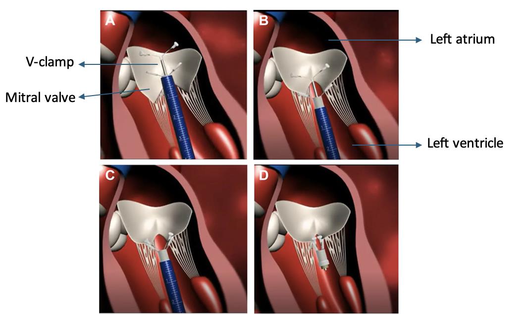
Transcatheter edge-to-edge repair (TEER) is performed using the V-clamp device. This is placed across the mitral valve to clip together the mitral valve leaflets and reduce the leaking. The steps are as follows:
- Access: Our cardiothoracic surgeons will make a small incision into the chest allowing visualisation of the heart. The interventional cardiologist will gain access to the heart using an introducer (small tube).
- Guidance: Advanced imaging using transoesophageal echocardiography, helps us visualize our equipment inside the heart and position the V-Clamp in the correct location.
- Repair: The V-clamp is tightened onto the valve, bringing the leaflet edges together and clamping them. This reduces the amount of valve leaking (figure 2).
- Recovery: The catheter is removed, the hole within the heart and the chest incision are closed and your dog is monitored closely after the procedure in ICU. Most dogs are up and walking the same day.
Complications:
The major risk of TEER is a partial detachment of the clamp from the mitral valve leaflets, which could lead to sudden death or severe heart failure signs that are almost always fatal. The highest risk of device detachment is within 48 hours of the procedure. Other less common risks include bleeding (which could be life threatening), blood clot events (thromboembolism), significant residual valve leakage, infection and anaesthesia related complications.
Planning a TEER referral to RVC
We are currently treating dogs with stage C degenerative mitral valve disease and monitoring those with less severe disease.
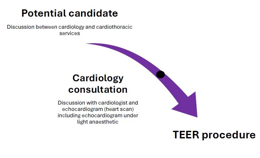
If you do not have a veterinary cardiologist but want to investigate whether TEER is a good option for your dog, please ask your primary care vet to make a referral to the QMHA cardiology service. We can then start the process with a face-to-face consultation and a specialist level heart scan (including 3D imaging).
If you have already seen a veterinary cardiologist, they will need to send us the images from your dog’s heart scan (echocardiogram). We will then discuss alongside our cardiothoracic surgery service whether they are likely to benefit from TEER, or if another treatment option should be considered. We will then organise a consultation with us to discuss TEER and perform a more detailed evaluation of the mitral valve using a special type of heart scan (transoesophageal echocardiogram) - this requires a short and light general anaesthesia. The information from this scan will help us carefully weigh up the benefits and risks of TEER, as well as plan for the procedure. After discussing the benefits and risks of the procedure, we will then provide a provisional TEER date.
If you have any questions regarding TEER, please email qmhacardiologyservice@rvc.ac.uk FAO TEER programme and a member of the cardiology service will respond within 72 hours.