24 hour contact: 01707 666297
Bone Scan (Scintigraphy)
Scintigraphy is one of the advanced imaging modalities used in horses. It uses radioactive tracers that allow the identification of changes in bone metabolism before they become visible on radiographs. It is ideally suited to diagnose for example hairline fractures very early on.
It allows identification of subtle problems in any area, but is especially useful in areas where clinical examination including diagnostic analgesia is often difficult, such as the pelvis and back.
The RVC's nuclear medicine suite is equipped with the only camera system in the world that has been developed specifically for horses, by the world’s leading manufacturer for high-end human systems. Unlike the second hand systems most other hospitals use, this system provides better image quality and shorter scanning times, resulting in the best possible service for your horse while keeping the time to a minimum.
How does it work?
- Nuclear Scintigraphy is used to image a tissue’s function and metabolic turnover.
- A radioactive tracer is injected into the animal, most commonly Technetium-phosphonates.
- This then distributes around the body and is taken up by areas with increased blood supply and metabolic activity.
- These areas emit Gamma rays, which are invisible to the human eye, but are detected by a gamma camera, which produces an image of the function, shape, size and position of the target organ (i.e. bone, thyroid etc.).
What does it show?
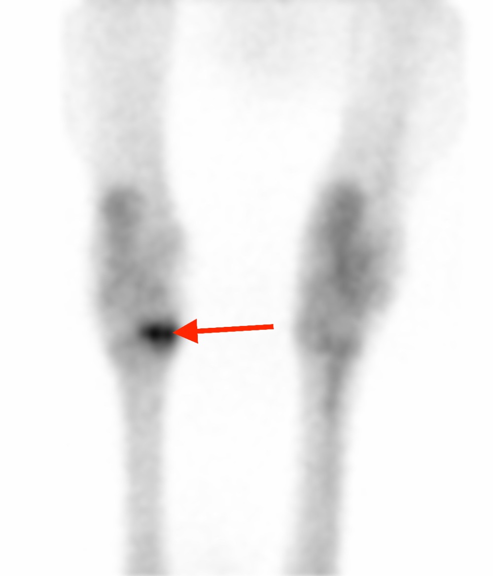
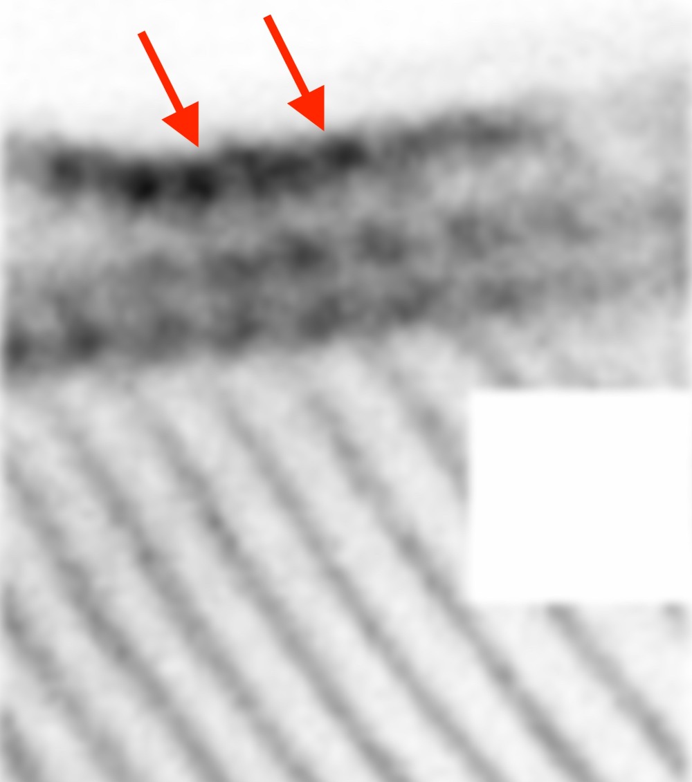
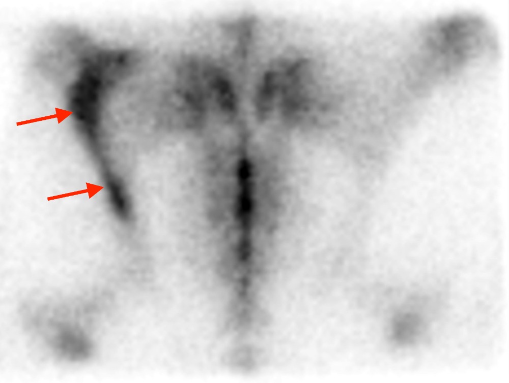
Areas of increased bone turn over are shown as 'hot spots' on images.
It is the most sensitive method for the detection of fractures, inflammation, and infection and it shows pathology earlier than any other method.
When do we use it in the horse?
- Horses where a fracture is suspected, but it is not visible on radiographs
- Back or pelvic problems
- Multi-limb lameness
- Horses that are intolerant of nerve blocks
- Obscure/intermittent lameness
- Poor performance

Bone scan image of the hocks taking from behind the horse. Note the marked increased uptake of the technetium (‘hot spot’) within the small tarsal bones. This horse showed marked osteoarthritic changes of the small tarsal joints on radiographs.

Lateral image of the back in a horse with ‘kissing spine’. Note the marked hotspots at the spinous processes of the thoracic spine (red arrows). Scintigraphy is especially helpful in these cases to decide if the changes are active and clinically significant or if they are chronic and do not cause pain.

Fracture of the ilium, one of the pelvic bones, visible as marked hotspot on the left side of the image (red arrows). This image was taken from the top of the horse. Such fractures are sometimes only diagnosed with the help of a bone scan.

